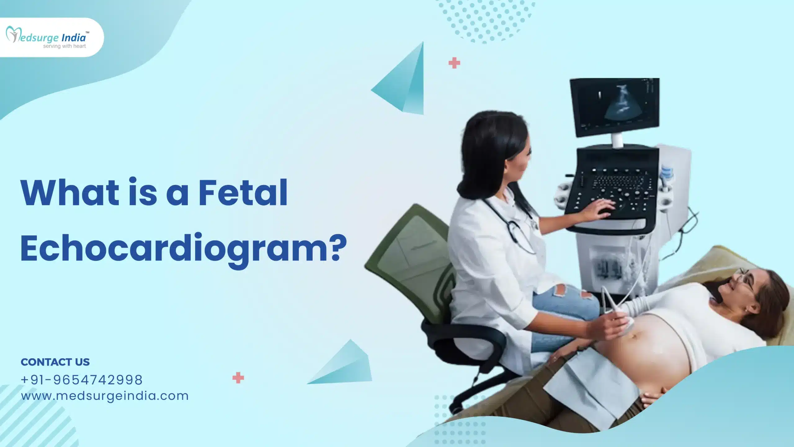
Fetal echo is short for fetal echocardiogram, this is an ultrasound test that doctors use to diagnose congenital heart disease (CHD). Congenital heart diseases, defined as diseases present at birth, affect about 1% of live births per year and include any abnormality of the heart. One type of CHD that is often diagnosed is termed ventricular septal defect abbreviated as VSD for short.
Understanding A Fetal Echo
In a fetal echocardiogram, the probe one uses is referred to as a transducer. It uses high-frequency sound waves to produce a picture of your stomach on the doctor’s computer monitor. These sound waves are at a frequency that goes through your skin and your baby’s skin to reflect off the heart structures.
Unlike other tests, which may involve some level of discomfort to the mother or the baby, this test does not in any way put the welfare of the baby in danger. It takes about 30 minutes to 2 hours because of the delicate and developing heart of the infant and because there are many things a doctor is searching for.
This fetal echocardiogram is almost similar to other ultrasound scans undertaken throughout pregnancy. It differs because the procedure is solely designated to view your baby’s heart without any other organs.
When Is Fetal Echocardiography Carried Out?
A fetal echocardiogram is not required in all pregnant women. In the majority of cases, the standard ultrasound will reveal the formation of all four walls of the baby’s heart.
An OB-GYN may suggest that you undergo this procedure if earlier tests were inconclusive or if the fetus had an irregular heartbeat.
It may also need this test if:
- expectant mother has the concern that an unborn child is predisposed to a heart disorder, or other condition.
- are suffering from heart diseases and 46 (50%) of them have a family history of heart disease.
- who have already had a child with a health complication such as heart disease.
- used drugs or alcohol during your pregnancy.
- used some types of drugs or received some medications that may lead to heart defects, for example, drugs used by epileptics, or drugs used for the treatment of acne.
- have other diseases, for instance, rubella, type 1 diabetes, systemic lupus erythematosus, or phenylketonuria.
A few OB-GYNs conduct this test. However, most of the time the test is done by a qualified ultrasound technician commonly referred to as an ultrasonographer. An experienced pediatric cardiologist will then go through the findings of the echocardiogram test.
How Is a Fetal Echocardiogram Done?
The test is done by a special ultrasound technician who has been specialized to take pictures of the fetal heart and these pictures are later read by a pediatric cardiologist who has specialized in fetal congenital heart disease. The fetal heart can only be briefly assessed during routine obstetric ultrasound and should only be performed in low-risk women. Nevertheless, the women, who have at least one of the mentioned risk factors, should undergo fetal echocardiography with a higher definition.
There are two ways to perform a fetal echocardiogram:
- Abdominal ultrasound: This is the most popular approach used to assess the state of the unborn baby’s heart. A gel is put on the woman’s stomach, a probe is then put on the stomach and pictures taken. This test is absolutely not invasive and is in no way dangerous to the fetus or the woman. The test usually lasts around 45 – 120 minutes if the fetal heart is complex.
- Endovaginal ultrasound: This method of ultrasound is done at the initial stages of pregnancy. A rather small ultrasound transducer is introduced into the vagina and positioned adjacent to the vagina’s back side. It is then possible to make photographs of the fetus’s heart.
Also read: Top 10 Gynecologists in India
What Happens During Fetal Echo?
Fetal echo is done by a pediatric cardiologist with special training in fetal cardiology, a maternal-fetal medicine specialist, an obstetrician or a radiologist, whichever best suits the case. In general, the steps include:
- An exam table is meant to made for you to lie on when the doctor is testing you. In all probability, you won’t need to change your clothes.
- The provider will place gel onto your abdomen.
- The provider will aim for an electronic device known as a transducer that emits sound waves.
- He or she will then manipulate the transducer in a bid to get the best pictures of the fetal heart. It can be a bit uncomfortable when the transducer is run on your belly.
- After the test is over the gel is washed off from the body part which has been used in carrying out the test.
At times in the course of pregnancy, an endo-vaginal echocardiogram is taken to check on the heartbeat of the developing fetus. The healthcare provider places a tiny echocardiogram probe into the vagina instead of placing it on the abdomen.
Also read: Top 10 Pediatric Cardiologists in India
What Will the Results Show?
Your health care professional will then explain the results of the test at your second appointment. If the test is normal, there were no issues noted as regards the cardiac function. You will carry on with the normal antenatal care and investigations as appropriate for pregnancy.
Abnormal results will inform you whether or not there is a matter with the functioning of the fetal heart or the development of it. It can also let you know if you have any irregular rhythms of the heart commonly known as arrhythmias. If any unusual values are present, you might have to get other tests done and in any case you would be recommended to see a doctor who deals with the specific disease.
In the course of even a normal study you will be advised that some heart problems are inevitable and incomprehensible. This is so because in circulation within the fetus unlike after birth. However, small interatrial communications between the lower chambers of the heart are particularly difficult to visualize and it is not always possible to obtain views of all the parts of the large blood vessels of the fetus’s heart.
It is very crucial to note that the diagnosis of heart defect has effects on the general well-being of the fetus. These health conditions could have potential consequences on the outcomes of the child and definitely contributes to some of the decisions you have to make about the pregnancy. Your cardiologist will let you know about whether you should be concerned with these other issues; however, when the concern is substantial, you will be referred to other members of the care team that collaborate with the pediatric cardiologist regularly.


