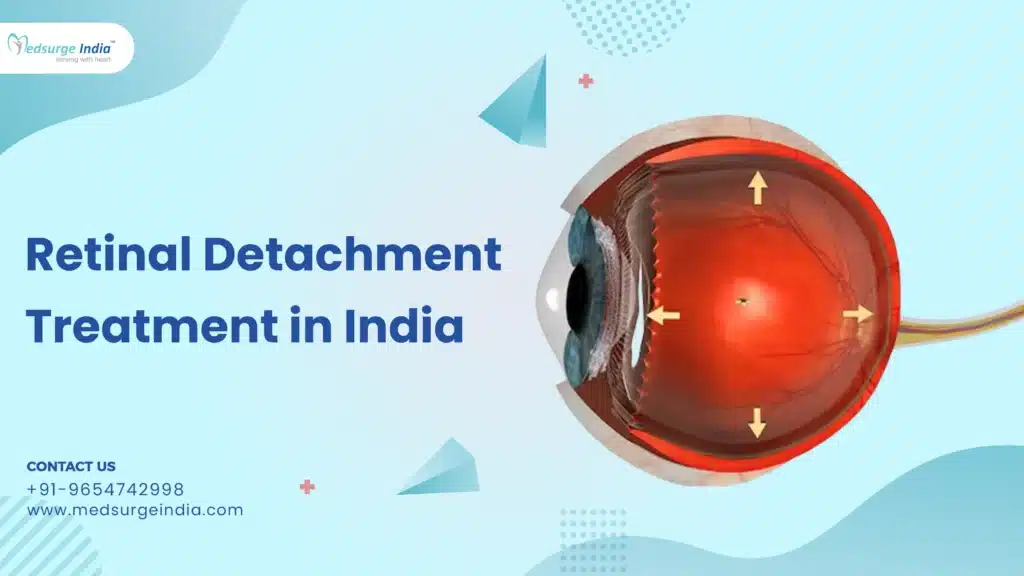
Retina Detachment Surgery Cost in India
Retinal Detachment Treatment Cost in India starts from 500 USD to 1500 USD. Alternatively, the cost is comparatively different from one patient to another because of one or the other factor. In minor cases of retinal tears or holes, the cost could range anywhere between 350 USD and 1350 USD based on the procedure.
Usually when healthy and normal eyes are being considered the possibility of developing retinal detachment would be 5 cases in every one hundred thousand individuals. For elderly people, this risk remains high; in people with severe myopia, it rises even higher. It also frequently develops as a postoperative infection following cataract surgery. It is however essential to note that if diagnosed early then can be well managed and treated through various techniques some of which are expensive.
In most cases, an ophthalmologist is bound to give such an estimate after a prognosis has been made based on the condition of the eyes.
Retinal Detachment Treatment in Different Cities in India
| Cities | Price Starting From |
| Delhi | 1100 USD |
| Mumbai | 1400 USD |
| Noida | 1250 USD |
| Gurgaon | 1200 USD |
| Chennai | 1500 USD |
| Kolkata | 950 USD |
| Bangalore | 1450 USD |
Factor That Can Affect the Cost of Retinal Detachment Treatment in India
- Treatment Type: Different treatments for retainal are tailored to address particular concerns. The complexity and nature of the chosen treatment significantly influence the overall cost.
- Location of the Hospital/Clinic: Variations in the cost of living and operational expenses across different cities and regions in India mean that the location of the clinic or hospital can substantially affect the total treatment costs.
- Treatment Duration: The financial burden may increase if the treatment requires several sessions, resulting in a higher cumulative expense for the patient.
- Technology and Equipment: The incorporation of cutting-edge technology and high-quality equipment in treatments can raise costs. Advanced methods often provide better outcomes, which can justify the elevated price.
- Severity of the Issue: The degree of the hair condition and the level of treatment required will affect the overall cost. More severe cases may necessitate additional sessions and products, leading to greater financial investment.
- Aftercare Requirements: Some treatments require aftercare, which may include medications, follow-up visits, or specialized hair care products. These additional costs should be included in the overall financial assessment.
Also, do keep in mind that the cost mentioned is only for the treatment as the total cost will depend on the final treatment after adding up all the factors that are mentioned above. Now let us get to know about the retinal detachment treatment and how it is treated.
What is Retinal Detachment?
Retinal detachment is a medical condition that refers to the detachment of a thin layer of tissue called the retina in the back of the eye.
Retinal detachment involves the physical separation of the cells in the retina from the underlying layer of blood vessels that supply oxygen and nutrients to the eye. If the retina is detached for a very long period without treatment, you are likely to lose eyesight in that particular eye permanently.
What Are the Types of Retinal Detachment?
There are three types of RD:
- Rhegmatogenous Retinal Detachment: A rhegmatogenous retinal detachment occurs when there is a tear or break in the retina that allows fluid to leak through into the sub-internal limiting membrane space.
- Exudative or Secondary Retinal Detachment: Exudative retinal detachment happens when inflammation takes place and it leads to the accumulation of the fluid beneath the retina without the formation of the hole or break.
- Tractional Retinal Detachment: A traction retinal detachment happens when fibrovascular tissue on the surface of the retina has pulled the retina from contact with its underlying tissues such as in the situation of a diabetic retinopathy case.
What Are the Symptoms of Retinal Detachment?
Retinal detachment is typically devoid of pain; however, it is often preceded by warning signs that may indicate its onset or progression. These signs include:
- The abrupt emergence of numerous floaters—small particles that appear to move across your visual field
- Flashes of light in one or both eyes (photopsia)
- Distorted or blurred vision
- A gradual decline in peripheral vision
- A shadow resembling a curtain that obscures part of your visual field.
How Is It Diagnosed?
The physician may employ the following diagnostic tests, instruments, and procedures to identify retinal detachment:
Optical coherence tomography (OCT): Typically, dilating eye drops will be administered prior to this imaging procedure. You will then position yourself in front of the OCT machine, resting your head on a support to ensure stability. The machine will scan your eye without making any physical contact.
Fundus imaging: Your healthcare provider may capture wide-angle photographs of your retina, usually after dilating your pupils for optimal visibility during the test.
Computed tomography (CT scan): This imaging technique integrates X-rays with computer technology and is generally utilized in cases involving a history of trauma or potential penetrating eye injuries.
Ocular ultrasound: The provider will carefully place a device against the front of your eye for scanning purposes. You will be asked to close your eyes while the provider applies gel to your eyelids. With your eyes shut, you will be instructed to move your eyeballs as the doctor conducts the scan using the same device.
How Is It Treated?
Depending on the condition, your eye care provider will advise on the most appropriate course of action. Depending on the extent of the eye problem, you might require multiple treatments for the best outcome. Treatments include:
- Laser (thermal) treatment or cryopexy: Both techniques are effective in repairing a retinal tear if identified at an early stage. These procedures are typically performed in a clinical setting.
- Pneumatic retinopexy: This method is particularly suitable for small and easily manageable tears. The physician introduces a small gas bubble into the vitreous gel, which exerts pressure on the upper portion of the retina, thereby closing the tear. Patients are required to maintain a specific head position for several days to ensure the bubble remains in the correct location.
- Scleral buckle: In this procedure, a silicone band, known as a buckle, is sutured around the sclera, the white part of the eye. This band exerts pressure toward the tear or detachment, facilitating the healing process. The buckle is not visible and remains permanently in place.
- Vitrectomy: This surgical intervention is necessary for larger tears or detachments. The surgeon removes the vitreous gel and substitutes it with either a gas bubble or oil. A vitrectomy may also necessitate that the patient maintains a specific head position for some time.
Approximately 80% to 90% of retinal surgeries yield successful outcomes; however, multiple procedures may be required. Recovery of vision can take several months, and some individuals may not fully regain their vision, particularly in more severe instances.
A detached retina will not heal independently. It is crucial to seek medical attention promptly to maximize the chances of preserving vision.
After the procedure
Following treatment for a detached retina, you may experience some discomfort, which can persist for several weeks. Your healthcare provider will discuss options for pain management and other relief methods. It is also essential to rest for a few weeks. Please consult your provider regarding the appropriate time to resume exercise, driving, and other regular activities.
Additional expectations post-surgery include:
- Eye patch: Adhere to the duration for which your provider recommends wearing the eye patch.
- Head position: If a bubble was introduced into your eye, it is crucial to follow your provider’s instructions regarding head positioning. They will specify the required position and the duration to maintain it to facilitate your eye’s healing process.
- Eye drops: Your provider will provide guidance on the proper use of eye drops to aid in your recovery.
- Improved vision: You can anticipate noticing improvements in your vision approximately four to six weeks after the surgery, although it may take several months to experience the full benefits.
Best Hospitals in India for Retinal Detachement Treatment
India is known to haves the best healthcare facilities in the world. The country is also known for their hospitality and the care for patients who come to India for treatment. Here is a list of the best top hospitals for retinal detachment treatment in India:
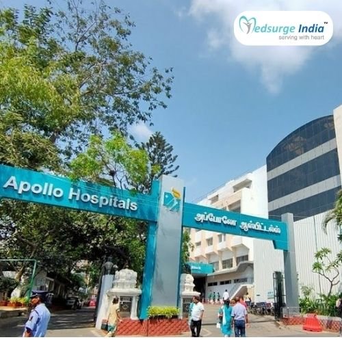
- Indraprastha Apollo Hospital
- Artemis Hospital, Gurgaon
- Manipal Hospital, New Delhi
- BLK-Max Super Speciality Hospital Delhi
- Fortis Memorial Research Institute, Gurugram
- Max Super Speciality Hospital, Saket (Max Saket)
- Medanta–The Medicity, Gurugram
Get Free Cost Estimation
The Most Important Frequently Asked Questions
Q: How Much Time Does Recovery Take?
A: It takes the patient at least a month to heal following the procedure. After the procedure, your eyes require careful attention. To fully recover, it’s also necessary to take the eye drops and prescription drugs as directed by the doctors for a predetermined amount of time.
Q: Are the Treatment’s Effects Long-Lasting?
A: The effects of detached retina surgery are essentially permanent after the patient has had it. But occasionally, patients may also require a second procedure.
Q: What Other Options Are There for the Treatment?
A: Surgical procedures are the most effective means of treating retinal detachment. Nonetheless, several nutrients and herbs are also recommended as other forms of treatment. Herbs including chamomile, bilberry, and pycnogenol can be used as an alternative therapy for the illness.
Q: for Whom Is the Treatment Appropriate?
A: People who experience light flashes, darkening of the peripheral vision, or a large number of new floaters or threads in their vision should get tested for retinal detachment very once. Due to the condition’s sudden development and lack of warning symptoms, it could quickly worsen. Those who have had cataract surgery, are severely nearsighted and have a family history of retinal detachment.
Top Hospitals for Retinal Detachment Treatment in India


Top Doctors for Ophthalmology

Dr. Sunaina Arora
Senior Consultant
Experience: 25 years of experience
Fortis Memorial Research Institute, Gurgaon
Gurgaon, India


Dr. Charu Khurana
Senior Consultant
Experience: 16 years of experience
Centre for Sight Eye Hospital, Rohini, Delhi
New Delhi, India

Dr. Gladys R Rodrigues
Senior Consultant
Experience: 19+ years of experience
KMC Hospital, Hampankatta, Mangaluru
Mangaluru, India

Dr. Srikanth R
Consultant
Experience: 14 years of experience
Apollo Spectra Hospital, Alwarpet, Chennai
Chennai, India

Dr. Svati Bansal
Consultant
Experience: 16 years of experience
Medanta – The Medicity, Gurgaon
Gurgaon, India


Dr. Chetana T Nayak
Consultant
Experience: 12 years of experience
BGS Gleneagles Global Hospitals, Bangalore
Bangalore, India

Dr. Bibhas H Shah
Consultant
Experience: 32 years of experience
Centre for Sight Eye Hospital, Akota, Vadodra
Vadodara, India

Dr. Atheeshwar Das
Consultant
Experience: 18 years of experience
Apollo Specialty Hospital, OMR
Chennai, India


Dr. Archita Agrawal Gupta
Consultant
Experience: 11 years of experience
Centre for Sight Eye Hospital, Akota, Vadodra
Vadodara, India

Dr. Arindam Deb
Consultant
Experience: 17 years of experience
Susrut Eye Foundation and Research Centre, Kolkata
Kolkata, India
Dr. Jayanti V
Experience: 32+ years of experience
Manipal Hospital, Mandi Mohalla, Mysore
Mysore, India

Dr. Sourabh Maheshwari
Consultant
Experience: 12 years of experience
Centre for Sight Eye Hospital Sapna Sangeeta
Indore, India

Dr. Sayan Das
Consultant
Experience: 19 years of experience
Susrut Eye Foundation and Research Centre, Kolkata
Kolkata, India

Dr. Sudhir Bhatia
Senior Consultant
Experience: 27 years of experience
Bharti Eye Hospital, New Delhi
New Delhi, India

Dr. Neha Bharti
Consultant
Experience: 9 years of experience
Bharti Eye Hospital, New Delhi
New Delhi, India