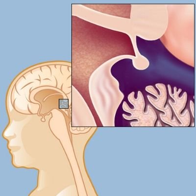
The Pineal Tumor is the growth of abnormal increases in the pineal gland or in the neighboring area. The pineal gland is a small, cone-shaped arrangement in the midline area of the brain situated in the bottom into the third ventricle. The pineal gland secretes neurotransmitters referred to as melatonin that modulates the circadian rhythm of your system. The receptor leads to various cells like parenchymal cells, endothelial cells, glial cells, and germ cells. The diversity of cells from the molecule presents tumors of distinct cellular sources in the cells. Pineal cell tumors are the ones that arise from the parenchymal cells included with the picture stimulus processing. The pineal tumors are termed as pineocytoma that is benign and slow and pineoblastomas that are malignant and competitive.
Symptoms
- Hydrocephalus due to increased pressure on the skull
- Disruption in hormonal production and release
- Nausea and Vomiting
- Headache
- Changes in vision
- The gradual loss of memory
Diagnosis
The indications of a brain tumor at the first stages aren’t routine and hard to diagnose as other diseases also cause symptoms that are similar. The neurologist can perform a neurological examination to detect the abnormality in vision, hearing, equilibrium, reflexes, and coordination. After the neural evaluation that the Physician might imply for additional tests like:
Computerized Tomography Scan (CT) – The CT scan creates two-dimensional brain images with high-power X-rays which is linked to a computer. The patient must lie on a movable table while shooting the images. A tracer dye will be injected through the I.V route after a few first CT scan images. The dye will create the tumors more visible, which frequently takes less than 10 min.
Magnetic Resonance Imaging Scan (MRI) – The scanning uses radio waves and magnetic fields to create the mind pictures. The patient needs to remain within a cylindrical device for 15 minutes to 60 minutes. MRI scans summarize the delicate tissues of the human body as well as bone that can be most commonly helpful in diagnosing brain tumors. A particular dye will be injected into the blood making tumors to differentiate from healthy tissue.
Head and Skull X-ray- Head X-rays will be done in order to detect alterations in the bone. Additionally, it finds a hydrocephalus condition connected with a brain tumor. Sometimes it can also detect the calcium deposits are connected with the tumor. Even though X-rays are routinely conducted, the test is less sensitive compared to other imaging tests.
Angiogram – A unique dye will be injected into the bloodstream which flows through blood vessels inside the brain to discover blood vessels in the vicinity of the tumor.
Other Tests – such as:
- Other tests like magnetic resonance spectroscopy (MRS), positron emission tomography (PET), single-photon emission computerized tomography (SPECT) scans can analyze the brain activity by exploring brain metabolism and chemistry.
- The tests combined with MRI scans will be of help to comprehend the purpose and brain activity but cannot be utilized in the main diagnosis of this tumor.
- The complete test to diagnose brain tumors is a biopsy. The biopsy may be required to collect a little portion of brain tissue like needle biopsy that may be carried out with the assistance of either CT scan or the component of the tissue removed because of part of the operation.
- The tissue investigation may discover the Sort of tumor, quality of tumor and Also Help Find out the treatment
- Cerebrospinal fluid evaluation to detect the existence of particular substances like Beta-HCG, AFP, etc.
- Blood evaluation ought to be asked to detect the mark from the blood.
Get Free Cost Estimation
Procedure
Treatment
Surgery
- Surgery is the first-line therapy for your pineal tumors. Even though it’s hard to remove a tumor in the pineal area of the brain however, the current advances in operation have made it a very powerful treatment option.
- The operation called craniotomy is performed by a neurosurgeon in which a part of the skull has been removed to access the tumor site and excised. If hydrocephalus is discovered shunt is put before the operation.
- The goal of surgery is to completely eliminate the tumor whilst maintaining the neighboring tissue. The benign pineal tumors will be treated after complete resection however, the cancerous tumors are hard for complete resection leading to subtotal resection that boosts the efficacy of adjuvant treatments.
Chemotherapy and Radiation
- In certain patients, tumor still remains after the operation, mostly the cancerous type. Sometimes, chemotherapy and radiotherapy are provided.
- The neurosurgeon will decide which treatment will be successful after the operation for complete treatment.
- The germinomas also referred to as germ cell tumors and pineoblastomas respond nicely to radiotherapy.
- The non-germinomatous and pineoblastomas are treated with chemotherapy. However, chemotherapy treatment for brain tumors isn’t well studied.
- Pineal cysts that are asymptomatic don’t require immediate treatment and are stored under observation.
The Most Important Frequently Asked Questions
Q: What are the symptoms of a Pineal Tumour?
A: The indicators of the pineal tumor are based on the size and location of the tumor. The most frequent symptoms are nausea, headache, numbness on the side of the face as well as migraines.
Q: What causes Pineal Tumour?
A: The primary source of pineal tumors isn’t known, the significant reasons are the strange genes, radiation, and chemical exposure. Individuals with inherited genes possess a higher risk for developing tumors like Li-Fraumeni syndrome, von Hippel-Lindau disorder, neurofibromatosis, and retinoblastoma. Individuals working at rubber and chemical industries can also be at risk for tumors. Previous radiation treatment for head and neck cancers makes it a risk factor for pineal tumors.
Q: How Pineal Tumours are treated?
A: Surgical removal of the tumour is the first alternative for a pineal tumor nevertheless chemotherapy and radiation also given in addition to the operation.
Q: What is the survival rate with Pineal Tumours?
A: The overall average survival period with pineal tumors is almost 5 decades and the survival rate with minimum complications following the treatment of up to 3 years is almost 85%.
Q: How common are the Pineal Tumours?
A: Pineal tumors develop in the area of the pineal gland. The gland is a small structure located deep within the brain that represents 1% of brain tumors and 3-8percent of the intracranial tumors mostly occurring in children.
Q: What is the difference between the pineal cyst and Pineal Tumour?
A: The pineal cyst is a generally non-malignant cyst from the pineal gland situated within the brain. The cysts seem as fluid-filled sacs at 1-4percent MRI scans and allegedly seen in 4-11percent of autopsies.
Q: How important is the pineal gland?
A: The pineal gland is a small cone-shaped gland that secretes various hormones and chemicals to get healthy and well-functioning processes. The hormone cortisol secreted from the pineal gland regulates the adrenal body rhythms.
Q: What are the risks of Pineal Tumour surgery?
A: The dangers of pineal tumor operation involves the formation of blood clots, swelling in the brain, infection in the wound site, an allergic response to anesthesia, memory issues, Impaired balance, vision and address, and coma.
Q: How the pineal tissue will be collected for a biopsy?
A: The tissue may be accumulated after the operation to remove the tumor or employing another needle biopsy procedure where only a tiny sample of tissue is obtained in the tumors that are found deep within the mind. The tissue is eliminated with the needle, and this is often directed by CT scanning.
Q: What is the follow-up after Pineal Tumour removal surgery?
A: Following the operation, chemotherapy and radiation may damage the hormonal creation of the human body. The hormonal imbalances may be treated with a referral to an endocrinologist.
Q: What are the eligibility requirements for a medical visa to India?
A: A medical visa to India needs a valid passport along with the treatment procedure accepted in a hospital accepted by the government of India with an expert panel of physicians. Two attendants might accompany the individual on another attendant visa with no more than 3 entries each year.
Top Hospitals for Pineal Tumor Treatment in India


Top Doctors for Neurology And Neurosurgery

Dr. Vinay Goyal
Director
Experience: 21 years of experience
Medanta – The Medicity, Gurgaon
Gurgaon, India


Dr. Rohit Gupta
Director
Experience: 13 years of experience
Fortis Escorts Hospital, Faridabad
Faridabad, India


Dr. Rahul Saha
Senior Consultant
Experience: 9+ years of experience
NH Rabindranath Tagore International Institute of Cardiac Sciences, Kolkata
Kolkata, India

Dr. Sameer Gupta
Director
Experience: 18 years of experience
Marengo Asia Hospitals, Faridabad
Faridabad, India

Dr. Priyank Patel
Consultant
Experience: 12 years of experience
Apollo Spectra Hospital, Mumbai
Mumbai, India

Dr. Bharat P Sarkar
Consultant
Experience: 10 years of experience
Manipal hospitals Life’s On, Whitefield
Bangalore, India


Dr. Srikanth K P
Consultant
Experience: 12 years of experience
Manipal hospitals Life’s On, Whitefield
Bangalore, India


Dr. P. R. Krishnan
Senior Consultant
Experience: 15 years of experience
Fortis Hospital, Bangalore ( Bannerghatta Road)
Bangalore, India


Dr. Rana Patir
Director , MBBS, MS, MCh
Experience: 24 years of experience
Fortis Memorial Research Institute
Gurgaon , India

Dr. Guruprasad Hosurkar
Senior Consultant
Experience: 15 years of experience
Manipal Hospital Formerly Columbia Asia Referral Hospital, Bangalore (Yeshwanthpur)
Bangalore, India


Dr. Vinit Suri
Consultant
Experience: 38 years of experience
Indraprastha Apollo Hospital New Delhi
New Delhi, India

Dr. Rajasekhar Reddy K
Consultant , MBBS, MS, MCh
Experience: 25 years of experience
Continental Hospitals, Hyderabad
Hyderabad, India

Dr. A.K. Roy
Consultant
Experience: 42+ years of experience
Manipal ( Old Airport Road) Hospital
Bangalore, India
Dr. Manoj Sharma
Head of Department
Experience: 36 years of experience
Fortis Hospital Delhi Shalimar Bagh
New Delhi, India